


Weekly Updates | April 7 | #7

Update #1 | The exciting world of RNA
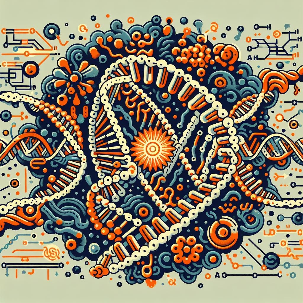
New findings from the Salk Institute provide evidence supporting the RNA World hypothesis, which suggests that RNA played a crucial role in early evolution. The study reveals an RNA enzyme that can accurately replicate and evolve RNA strands, offering insights into the origins and complexity of life.
Key points:
The RNA World hypothesis proposes that RNA molecules ruled the early Earth and established the dynamics of evolution.
Scientists at the Salk Institute have discovered an RNA enzyme that can make accurate copies of other RNA strands and allow new variants to emerge over time.
The findings suggest that evolution may have occurred on a molecular scale in RNA before the emergence of cells and DNA.
The research brings scientists one step closer to synthesizing RNA-based life in the laboratory.
Highlights:
The study provides strong evidence supporting the RNA World hypothesis and the role of RNA in early evolution.
The RNA enzyme developed by the researchers can accurately replicate and evolve RNA strands, suggesting the potential for RNA to support Darwinian evolution.
These findings have implications for understanding the origins and complexity of life and could pave the way for the synthesis of RNA-based life in the laboratory.
The research also raises questions about the conditions that supported RNA evolution and the potential for new functions to emerge in the early stages of life.
References:
“RNA-catalyzed evolution of catalytic RNA” by Nikolaos Papastavrou, David P. Horning and Gerald F. Joyce, 4 March 2024, Proceedings of the National Academy of Sciences. DOI: 10.1073/pnas.2321592121
Update #2 | The Most powerful MRI scanner
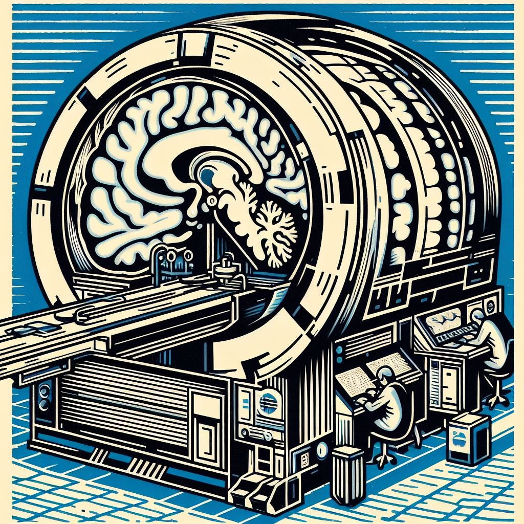
The world's most powerful MRI scanner, named "Iseult," has produced its first images of the human brain. The scanner, developed by researchers at France's Atomic Energy Commission (CEA), is capable of scanning images with 10 times more precision than conventional MRI machines commonly used in hospitals. The high level of precision is expected to provide a better understanding of brain functioning, as well as neurodegenerative and psychiatric diseases. The scanner's power could also help researchers study conditions like Parkinson's, Alzheimer's, depression, and schizophrenia, and improve detection and treatment methods.
Key points:
The world's most powerful MRI scanner, called "Iseult," has generated its first images of the human brain.
The scanner, located in France, is capable of scanning images with 10 times more precision than traditional MRI machines.
The scanner's high level of precision is expected to enhance understanding of brain anatomy and functioning, as well as neurodegenerative and psychiatric diseases.
Researchers hope the scanner's power can shed light on conditions like Parkinson's, Alzheimer's, depression, and schizophrenia.
The machine is not intended for clinical diagnosis at this point, but the knowledge gained from it can be used in hospitals.
Highlights:
The MRI scanner, "Iseult," is located in the Plateau de Saclay area in France and has a magnetic field strength of 11.7 teslas.
The scanner has already provided images with unprecedented precision, allowing for the visualization of tiny vessels that feed the cerebral cortex and details of the cerebellum.
The machine's precision could help researchers better understand the relationship between brain structure and cognitive functions.
Scientists hope the scanner can contribute to early detection and improved treatment of brain pathologies.
The scanner could also help researchers map how specific drugs distribute through the brain, potentially aiding in personalized treatment approaches.
Regular patients will not have access to the scanner for several years, as it is primarily for research purposes.
References:
Update #3 | New enzymes to treat autoimmune conditions
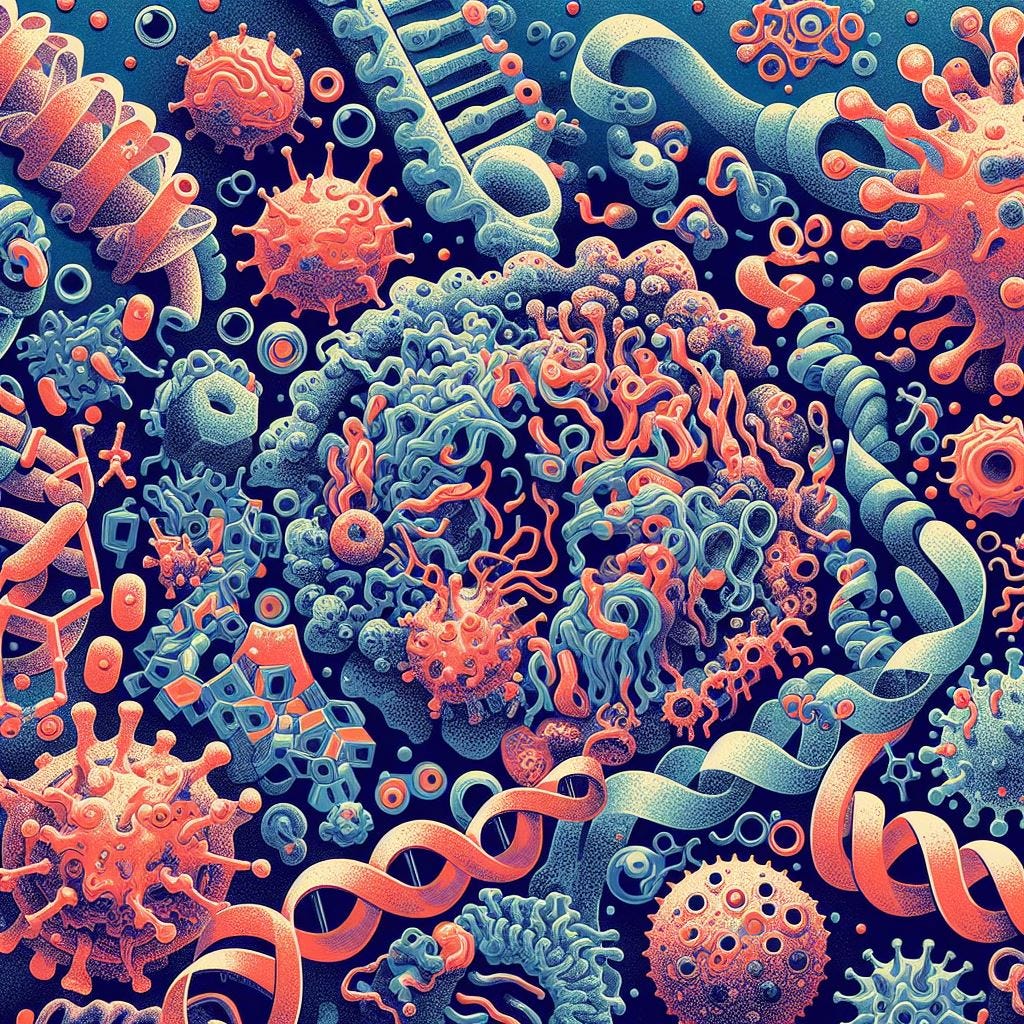
In a new study published in Structure, scientists from The Scripps Research Institute have presented the previously undescribed structures of two nucleic acid-degrading enzymes, PLD3 and PLD4. These enzymes play a role in clearing nucleic acids from a cell's cytoplasm and malfunctioning can lead to autoimmune and inflammatory diseases. Understanding the structures and molecular details of these enzymes is crucial for designing therapies for related diseases such as lupus erythematosus, rheumatoid arthritis, and Alzheimer's disease.
Key Points:
Scientists at The Scripps Research Institute have determined the structures of two nucleic acid-degrading enzymes, PLD3 and PLD4, which are essential for clearing nucleic acids from a cell's cytoplasm.
Dysfunction of these enzymes can lead to autoimmune and inflammatory diseases, making it important to understand their structures and functions for the development of therapeutic interventions.
The researchers used X-ray crystallography to build atomic-scale models of these enzymes, allowing for a detailed examination of their structures in different states.
The study revealed that PLD3 and PLD4 have structurally similar mechanisms for degrading nucleic acids.
The researchers also found a previously unknown function for PLD4, which showed phosphatase activity in addition to its nucleic acid-degrading capabilities.
Further investigations will focus on inhibiting overactive enzymes and potentially replacing non-functional versions of the enzymes.
Highlights:
The structures of nucleic acid-degrading enzymes PLD3 and PLD4 have been determined, providing insights into their mechanisms and function.
Dysfunction of these enzymes can lead to autoimmune and inflammatory diseases.
The research opens up possibilities for designing therapeutic interventions for related diseases.
The study also revealed a previously unknown function of PLD4, which has phosphatase activity in addition to its role in nucleic acid degradation.
Further investigations will focus on inhibiting overactive enzymes and potentially replacing non-functional versions.
References:
https://phys.org/news/2024-04-unheard-biology-enzyme-reveal-disease.html
Meng Yuan et al, Structural and mechanistic insights into disease-associated endolysosomal exonucleases PLD3 and PLD4, Structure (2024). DOI: 10.1016/j.str.2024.02.019
Update #4 | The anti-aging Tardigrades

A study from the University of Wyoming suggests that proteins found in tardigrades, also known as water bears, can slow down molecular processes in human cells. These proteins, which allow tardigrades to enter a state of suspended animation, or biostasis, could potentially be used to slow down the aging process and develop storage technologies for human cells. The study also found that the protein-induced gels dissolve away in human cells once the external stress is relieved.
Key points:
Certain proteins found in tardigrades can slow down molecular processes in human cells, potentially aiding in slowing the aging process.
Tardigrades can enter a state of suspended animation or biostasis, allowing them to survive extreme temperatures and radiation.
When these tardigrade proteins are introduced into human cells, they form gels and slow down metabolism.
Human cells with these proteins become more resistant to stresses, similar to tardigrades' abilities.
The protein-induced gels dissolve in human cells once the external stress is relieved.
Exploring the biomedical applications of tardigrade abilities has been a recent topic of study.
Highlights:
Tardigrades, also known as water bears, are microscopic organisms that can thrive in harsh conditions, including outer space.
Tardigrades have the ability to survive extreme temperatures ranging from -328 degrees Fahrenheit to over 300 degrees Fahrenheit.
Tardigrades can enter a state of suspended animation, allowing them to survive extreme conditions.
The proteins that enable tardigrades to enter biostasis form gels in their cells, slowing down their metabolism.
Introducing these tardigrade proteins into human cells can also slow down metabolism and make them more resistant to stresses.
Tardigrade proteins could potentially be used to slow down the aging process and develop storage technologies for human cells.
Biomedical applications of tardigrade abilities have been previously explored, including the creation of pharmaceuticals for treating hemophilia.
References:
Update #5 | Airborne Human DNA in Forensics

Airborne human DNA has the potential to be used in forensic analysis of crime scenes that have been wiped clean of fingerprints and trace evidence, according to a new study. Researchers found that human DNA can be found in the air and settle on surfaces, including air-conditioning units. This discovery opens up new avenues for gathering evidence and identifying individuals present at a crime scene.
Key points:
Offenders cannot completely prevent their DNA from being released into the environment.
Human DNA can be collected from the air and on the surfaces of air-conditioning units.
Environmental DNA (eDNA) and environmental RNA (eRNA) shed from sources such as skin or saliva can be detected in soil, ice, air, and water.
Air samples are likely to represent more recent occupation, while surface samples represent prior occupation.
Airborne DNA can be used to prove if someone has been in a room, even if they wore gloves or wiped surfaces clean.
Highlights:
Airborne DNA can be used in forensic analysis to gather evidence at crime scenes.
Potential to identify both usual users and visitors of a room.
Follow-up studies are recommended to determine the best location for air collection devices and the appropriate time to test for DNA after a crime.
References:
Update #6 | Vegan Leather

Scientists at Imperial College London have genetically engineered bacteria to produce vegan leather that is plastic-free and self-dyeing. The researchers modified genes in bacteria species to produce sheets of bacterial cellulose and the dark black pigment, eumelanin. The bacterial cellulose is grown around a mold shaped like a shoe to create a conventional upper. This new process has the potential to expand into different colors and materials, and it offers a sustainable and environmentally friendly alternative to traditional leather production methods.
Key points:
Scientists at Imperial College London have genetically engineered bacteria to grow animal- and plastic-free leather that can dye itself.
The researchers modified the genes of bacteria species to produce sheets of bacterial cellulose and the dark black pigment, eumelanin.
The bacterial cellulose was cultivated around a mold shaped like a shoe to create a conventional upper.
This new process offers a sustainable and environmentally friendly alternative to traditional leather production, as it requires fewer carbon emissions, water, land use, and time compared to farming cows for leather.
The bacteria can be engineered using genes from other microbes to produce colors in response to blue light.
The researchers are experimenting with various colored pigments that the material-growing microbes can produce.
Highlights:
Scientists at Imperial College London have genetically engineered bacteria to grow plastic-free, self-dyeing vegan leather.
The process involves modifying the genes of bacteria species to produce sheets of bacterial cellulose and the dark black pigment, eumelanin.
The bacterial cellulose is grown around a mold shaped like a shoe to create a traditional upper.
This sustainable and environmentally friendly alternative to traditional leather production has the potential to expand into different colors and materials.